Use Case
Precision Anatomic Models
The medical world has long been a fan of additive manufacturing, and with good reason.
By using various medical imaging techniques, users are able to convert this data into digital models, which can then be 3D printed for a whole range of medical applications.
A group of researchers from the San Francisco VA Health Care System and the University of California San Francisco (UCSF) has been using the technology to assist with education for doctors, and also for the planning of surgeries. Read on to know more.
Dr. Alexis Dang M.D is one of the researchers from the Department of Orthopaedic Surgery at the University of California, and much of his work has been focused on using additive manufacturing to improve the efficiency of orthopedic surgery. Since 2004, Dang has been an advocate of 3D printing across his campus, and is the co-founder of Edge Labs and also the UCSF CA3D+ project which focused on AM and AR/VR applications in medicine.

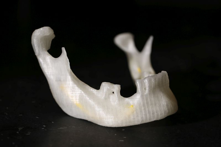
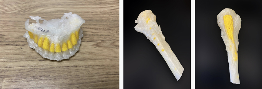
Much of the work at Edge Labs focuses on the design and manufacture of Precision Anatomic Models (dubbed as “PAMs” by the team) which are used for clinical decision making and surgical planning.
Having a tactile model of a part that one is about to operate on allows surgeons to envision their upcoming procedure visually and physically. Sure, a digital 3D model offers some outlook, and doctors have long used 3D visualization to plan their surgeries. But surgery is a highly tactile thing, indeed surgeons are renowned for their super dexterity.
And having a physical model of a broken bone in their hands before they start work offers an entire new, and critical dimension to their planning.

And naturally, being a university, the team has found use for the 3D printed bones in teaching. With their extruded polymer printers they print their broken and deformed bone models with PETG and PLA to educate doctors on the various bone issues.
There is another category of person who can also benefit from these precision anatomical models, and that is the patient. Now, thanks to these tactile demonstrations, doctors can use them for patient engagement, showing patients details in their own bones that could not be expressed simply via an X-ray. It adds a whole new dimension to the concept of “informed consent” with a patient.
Of course, bones can be pretty big, and on smaller printers can take a long time, so in order to meet demands, they invested in printers with higher flowing extruders and larger print beds.
In particular, they opted for the Craftbot Flow IDEX XL, which in addition to higher material deposition rates also features dual independent extruders, which the researchers use for printing objects in parallel, and also in dual colours and materials. The machine uses a CoreXY configuration to move the extruders about their axes.
Higher Throughput/Lower Costs
By using larger, faster machines that can also print parallel parts with the dual extruders, machine time becomes more productive by default. More parts are printed, and value is recognized quicker. These factors alone reduce the cost per part.
In addition, the Craftbot uses open source/3rd party filaments, so the researchers are not restricted to one vendor.
According to Dang, these factors and the open source filaments are allowing the team to create parts at one order of magnitude cheaper than those made on a 3D printer using proprietary (or DRM’d!) filaments.
This all means that Dang and the researchers can provide rapid services for medical professionals at a low cost. The Edge Labs website states that they are driven by stakeholders from the frontline, and so having a fast and cost effective solution is beneficial in keeping those stakeholders happy.
If you would like to read more about the work that Dr. Dang is doing, you can read more at the Edge Labs website, or indeed if you are a stakeholder and would like to order some parts for your own patient engagement or surgical planning, you can fill in the form on their homepage and request some bones be delivered straight to your door.


